Menu

Microstructural & Compositional Characterization

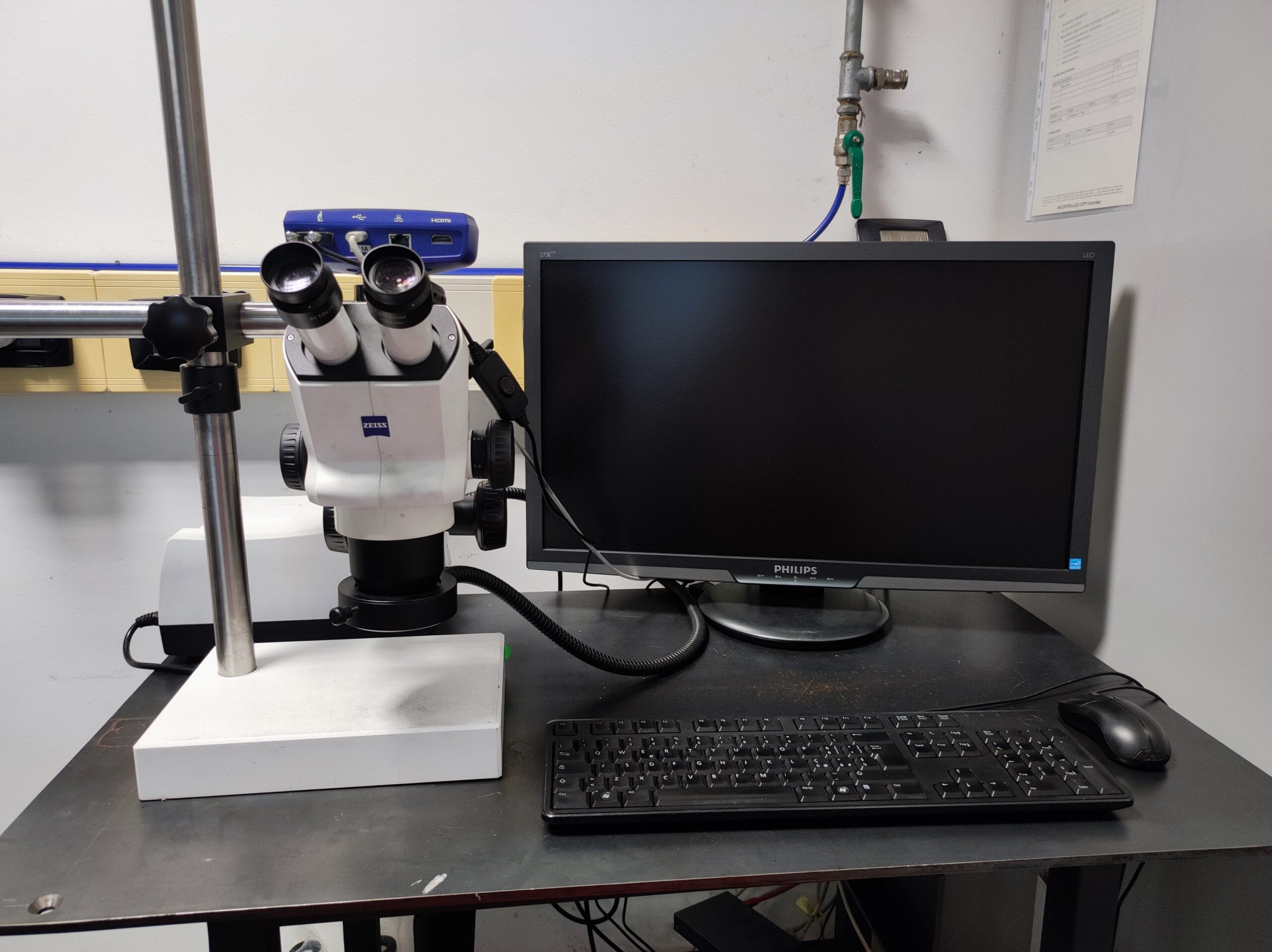
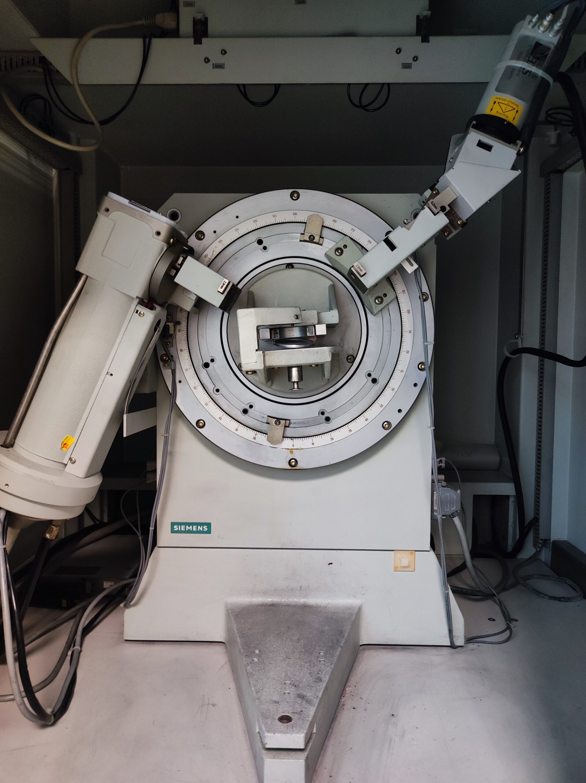
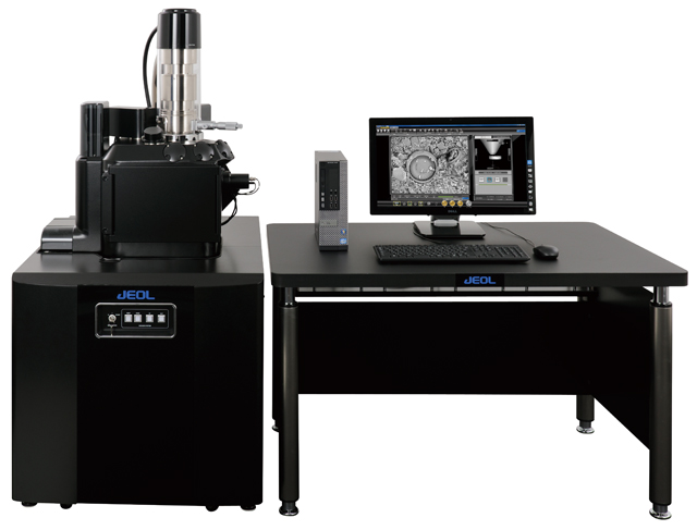
Microstructural and compositional characterization provides essential insight into material performance at a fundamental level. Advanced microscopy and analytical techniques allow detailed evaluation of grain size, phase distribution, interfaces, defects, and porosity within ceramic and composite systems.

Complementary chemical analysis ensures accurate identification and quantification of elements and phases, supporting control of purity, homogeneity, and formulation. The integration of structural and compositional data enables a clear understanding of structure–property relationships, supporting materials development, quality control, and failure analysis.

stereomicroscopio

Microscopy

XRD

X-ray Powder Diffraction

Close

Microscopy

Optical and electron microscopy are fundamental techniques for material analysis. Optical microscopy uses visible light and lenses to observe samples at magnifications of up to about 1000x, making it useful for rapid analysis of microstructures, defects, or porosity.

On the other hand, electron microscopy employs an electron beam and provides much higher resolutions (down to the nanometric level). Both are key tools in advanced research and development.

Stereomicroscope

stereomicroscopio
Eyepieces 10x/23 Br. Foc.
Front optics from 0.63x to 5.0x
Ring illuminator and Double spot illuminator
Polarization filter set S, consisting of fixed analyzer and rotary, annular polarizer
Microscopy Camera Axiocam 208 color

Electronic microscope SEM (JEOL JSM IT300LA)

Accelerating voltage> 300V
Magnificationup to 300,000x
Resolution3.0nm at 30kV and 15nm at 1kV
Imagesecondary and backscattered electron
Microanalysis with Energy Dispersive X-ray Spectroscopy (EDX)

Close

X-ray Powder Diffraction

X-ray powder diffraction (XRD) is an analytical technique used to identify the crystalline structure of materials. When a powder sample is irradiated with X-rays, they are diffracted at specific angles, which are related to the crystal lattice of the material.

By analyzing the resulting diffraction pattern, it is possible to determine the crystalline phases present, calculate the crystal size, assess internal strain, and estimate the degree of crystallinity

X-Ray diffractometer

XRD
GeometryBragg-Brentano
Reproducibility±0.0005°
Monochromatorgraphite
AnodeCopper
DetectorNaI (Tl), with reading speed of 105imp./s